,Hue:lla, De. La,MA>No De,, Un Ni(Ñ@) Des.Pue;De JugAr,Se Tra>NZ For.MA en UnI.VErZo, De MI:;Cro -ORGAniS,MOZ Kul;TiVA-DoZ

Screen Shot 2015-06-09 at 11.16.12 AM

As a living organism, the human body is home to millions of microbial life forms and bacteria. Without microscopic vision, we can’t see them unless aided by technology. If you’re the squeamish sort, this is probably for the best.

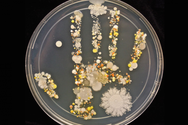
The handprint in the petri dish above is causing quite a stir on the Internet and it belongs to microbiology lab technician Tasha Sturm’s 8-year-old son.

“It’s partly to show that there are microbes everywhere,” said Sturm to TODAY.

Posted on Microbe World, the print shows the different growths cultivated from his hand after playing outdoors. Allowed to incubate for several days, there are yeasts, fungi and bacteria.

Sturm will conduct further tests to determine what exactly the various growths are. She believes the large white circle in the bottom-right corner (close-up shown below) to be Bacillus, which is often found in dirt. She also notes that the white spots may be Staphylococcus and the yellow and orange spots could be yeast.


Close-up of the round blob in the bottom-right corner of the handprint / Tasha Sturm

Sturm regularly engages her kids in science experiments at home, which has led to some interesting investigations, sometimes including the family dog. After petting the dog, her son did the same process of placing his hand in a sterile petri dish and incubating the dish at body temperature for a day, and then room temperature for nearly a week. His reaction to the results: “He said, ‘That’s cool.’ And then my daughter said, ‘Let’s do the dog’s nose, let’s do the paw, let’s do the cat’s tail,'” Sturm recalled.

It’s important to remember that the vast majority of this organisms will be harmless, or even beneficial to human health. We are constantly coated in a variety of different microorganisms, no matter how clean you are, and our skin does a great job of keeping out the nasty ones.

Tar-Di/Grade Meet PAr:Ty:CiPIo ::: .. Wa-TEr Be:Ar Ge:Nes aR3 · Pri:MAri-Ly Of. BakTE:RiaL (O)RiGin

tardigrade-1024x795

Source:::: http://news.meta.com/2015/11/23/waterbear/

PNAS: The tardigrade (water bear), the only animal that can survive in the vacuum of space, has the most foreign DNA of any animal.

Environment & Ecology, Genetics & Genomics – November 23rd, 2015 –

The tardigrade, also known as the water bear, is renowned for many reasons. The nearly indestructible micro-organism is known to have the capacity to survive extreme temperatures (-272C to 151C), and is the only animal able to survive in the vacuum of space.

Today, with the publication of its genome in PNAS, the humble water bear can add another item to its exhaustive list of superlatives. Sequencing of the genome, performed by a team of researchers at the University of North Carolina at Chapel Hill, has revealed that a massive portion of the tiny organism’s genome is of foreign origin. Indeed, nearly 17.5% of the water bear’s genome is comprised of foreign DNA, translating to a genetic complement of approximately 6,000 genes. These genes are primarily of bacterial origin, though genes from fungi and plants have also been identified.

Horizontal gene transfer, defined as the shifting of genetic material materially (thus horizontally) between organisms is widespread in the microscopic world. In humans, however, the process does occur, but in a limited fashion, and via transposons and viruses. Other microscopic animals are also known to have large complements of foreign genes.

Until today, the tiny rotifer was believed to have the greatest complement of foreign DNA of any microscopic organism. Surprisingly, that genetic complement constitutes only half of what has been identified in the newly published tardigrade genome.

The authors of the newly published work have proposed a method by which this extremely extensive gene transfer may have occurred. Tardigrades have long been known to undergo, and survive, the process of desiccation (extreme drying out). The authors therefore postulated that during this drying out process and the subsequent rehydration, the tardigrade’s genome may have undergone significant sheering and breakage, resulting in a general loss of integrity and leakiness experienced by the water bear’s nucleus. In turn, this compromised nuclear integrity may have enabled foreign genetic material to readily integrate the genome, in much the same way as scientists perform gene transfer through the process of electroporation.

For now, the tardigrade has a dual claim to fame, being the only known animal to survive the vacuum of space, and being the animal with the largest genetic complement. Only with the study of other micro-organisms will we be able to validate if the humble tardigrade maintains its two, current, great claims to fame.

Source: Thomas C. Boothby, Jennifer R. Tenlen, Frank W. Smith, Jeremy R. Wang, Kiera A. Patanella, Erin Osborne Nishimura, Sophia C. Tintori, Qing Li, Corbin D. Jones, Mark Yandell, David N. Messina, Jarret Glasscock, and Bob Goldstein Evidence for extensive horizontal gene transfer from the draft genome of a tardigrade PNAS 2015 ; published ahead of print November 23, 2015, doi:10.1073/pnas.1510461112

QuanTuM, (tELe)TRaNS:pOR:TATion Of Micro:OrgA,NisimS

1-physicistsprBAkteria

So:uR_Ce : http://phys.org/news/2016-01-physicists-scheme-teleport-memory.html

Physicists propose the first scheme to teleport the memory of an organism

January 14, 2016
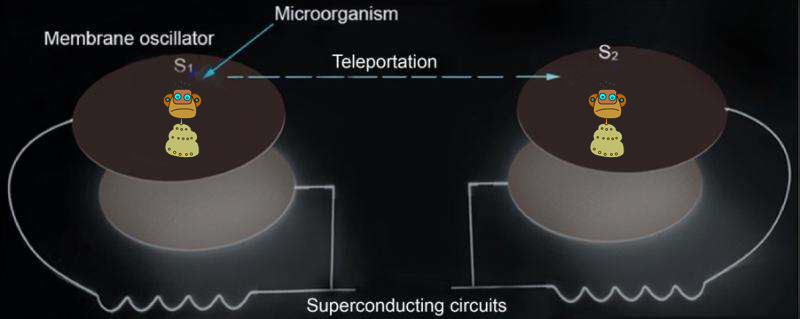
Quantum teleportation between two microorganisms is shown. The internal state (an electron spin) or the center-of-mass motion state of a microorganism on an electromechanical oscillator can be teleported to a remote microorganism on another …more

In “Star Trek,” a transporter can teleport a person from one location to a remote location without actually making the journey along the way. Such a transporter has fascinated many people. Quantum teleportation shares several features of the transporter and is one of the most important protocols in quantum information. In a recent study, Prof. Tongcang Li at Purdue University and Dr. Zhang-qi Yin at Tsinghua University proposed the first scheme to use electromechanical oscillators and superconducting circuits to teleport the internal quantum state (memory) and center-of-mass motion state of a microorganism. They also proposed a scheme to create a Schrödinger’s cat state in which a microorganism can be in two places at the same time. This is an important step toward potentially teleporting an organism in future.

In 1935, Erwin Schrödinger proposed a famous thought experiment to prepare a cat in a superposition of both alive and dead states. The possibility of an organism to be in a superposition state dramatically reveals the profound consequences of mechanics, and has attracted broad interests. Physicists have made great efforts over many decades to investigate macroscopic quantum phenomena. To date, matter-wave interference of electrons, atoms, and molecules (such as C60) have been observed. Recently, quantum ground state cooling and the creation of superposition states of mechanical oscillators have been realized. For example, a group in Colorado, U.S. has cooled the vibration of a 15-micrometer-diameter aluminum membrane to quantum ground state, and entangled its motion with microwave photons. However, the quantum superposition of an entire organism has not been realized. Meanwhile, there have been many breakthroughs in since its first experimental realization in 1997 with a single photon. Besides photons, quantum teleportation with atoms, ions, and superconducting circuits have been demonstrated. In 2015, a group at University of Science and Technology of China demonstrated the quantum teleportation of multiple degrees of freedom of a single photon. However, existing experiments are still far away from teleporting an organism or the state of an organism.

In a recent study, Tongcang Li and Zhang-qi Yin propose to put a bacterium on top of an electromechanical membrane oscillator integrated with a superconducting circuit to prepare the quantum superposition state of a microorganism and teleport its quantum state. A microorganism with a mass much smaller than the mass of the electromechanical membrane will not significantly affect the quality factor of the membrane and can be cooled to the quantum together with the membrane. Quantum superposition and teleportation of its center-of-mass motion state can be realized with the help of superconducting microwave circuits. With a strong magnetic field gradient, the internal states of a microorganism, such as the electron spin of a glycine radical, can be entangled with its center-of-mass motion and be teleported to a remote microorganism. Since internal states of an organism contain information, this proposal provides a scheme for teleporting information or memories between two remote organisms.

The proposed setup is also a quantum-limited magnetic resonance force microscope. It can not only detect the existence of single electron spins (associated with protein defects or DNA defects) like conventional MRFM, but can also coherently manipulate and detect the quantum states of electron spins. It enables some isolated electron spins that could not be read out with optical or electrical methods to be used as quantum memory for quantum information.

Li says, “We propose a straightforward method to put a microorganism in two places at the same time, and provide a scheme to teleport the of a microorganism. I hope our unconventional work will inspire more people to think seriously about quantum teleportation of a microorganism and its potential applications in the future.” Yin says “Our work also provides insights for future studies about the effects of biochemical reactions in the wave function collapses of states of an organism.”

Read more at: http://phys.org/news/2016-01-physicists-scheme-teleport-memory.html#jCp

Ci;ENTiFi;Koz Vou:YE;Ri.Ztaz , VieN:Do PoRNO Eu:KA-RyO_TiKO El SeX.O en :LoS., Or;GA;NIzMoz _ MuL:Ti.Ce.Lu*LA.Rez

sexamongeukaPiZtilo

sou:RCe : 

Sex among eukaryotes is far more common than once believed

July 28, 2015 by Christopher Packham report
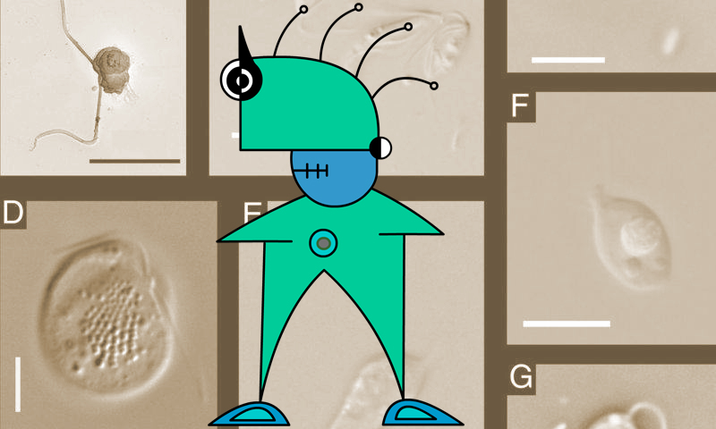
Representatives of deep eukaryotic lineages without published evidence for sex thus far. (A) Picomonas judraskeda (Picozoa). (B) Andalucia incarcerata and another, thus far undescribed jakobid (Jakobida). (C) Ancyromonas sigmoides …more

(Phys.org)—For a long time, biologists have considered sex to be an inherent trait of multicellular life, while microbial eukaryotes were considered to be either optionally sexual or purely clonal. From this perspective, clonality in eukaryotes is seen as exceptional. However, a group of researchers Europe and Canada have recently published a paper examining this broad distinction more closely, and have suggested that it appears to stem from an improper comparison of unicellular and multicellular species.

The paper, published in the Proceedings of the National Academy of Sciences, points out that in is simply clonal cell propagation among physically linked cells. “Hence,” the researchers write, “from the perspective of cell lineage, sex in multicellular organisms is as episodic as it is in facultatively sexual unicellular eukaryotes.” The authors’ emphatic conclusion is that “sex is a ubiquitous, ancient, and inherent attribute of eukaryotic life.”

Notably, the paper emphasizes that zoologists would be aghast at the absence of observed sex, while microbiologists are far more receptive to the lack of sex in protists. Many protist groups, including ciliates and green algae, propagate via sex, but direct observation of those processes is lacking for the vast majority. Indeed, there are entire lineages of protists for which no evidence of sex processes exists. However, the authors screened scientific literature to find individual “signs of sex” in eukaryotic lineages, including physical observation of cell fusion or nuclear fusion, genetic evidence of meiosis or recombination, or changes in ploidy levels over the life cycle.

Among the individuals screened, Jakobida, Glaucophyta, and Malawaimonadida—putatively asexual eukaryotes— were all found to contain genes involved in gamete fusion and/or . The authors suggest that sex among unicellular eukaryotes is likely to be far more common than currently believed, and the lack of evidence of sexual propagation attributable to the difficulty of microbiological observation. Highlighting this difficulty, they point out a famous example of a particular type of algae with two morphologically different stages, which had been wrongly considered to be two separate species. What we don’t know about protist life forms still vastly outweighs what we’ve discovered. “…(W)e still have a tendency to underestimate how widespread sexual practices are in the different eukaryotic groups,” the authors write.

Further, genome sequencing now supports the fundamentally sexual nature of eukaryotes. The authors cite numerous examples of putatively asexual eukaryotes found to express genetic traits associated with sex propagation. Giardia intestinalis was assumed to be asexual until genomic inspection revealed allelic differences indicative of sex. “The list of eukaryotic species that lack strong direct evidence for meiotic sex, but that seem sexual, as suggested by the presence of these meiosis-associated genes, is growing longer and longer,” the authors write.

There are numerous adaptive benefits to sex: It creates genetic variation, repairs DNA breaks, and prevents the accumulation of disadvantageous mutations. Sexual reproduction is also associated with species survival during adverse periods. Because of these advantages, the authors suggest, even asexual species overwhelmingly retain the option for meiotic sex propagation, even despite some of the disadvantages of sex for protists.

The paper goes on to speculate on the possibility that the evolution of meiotic sex was a defensive response to DNA-damaging effects of reactive oxygen species, and considers the possible influence of endosymbiotic organisms like chloroplasts and mitochondria on the evolution of sex.

Explore further: Researchers Present New Sex Evolution Theory

More information: “Sex is a ubiquitous, ancient, and inherent attribute of eukaryotic life.” PNAS 2015 112 (29) 8827-8834; published ahead of print July 21, 2015, DOI: 10.1073/pnas.1501725112

Abstract
Sexual reproduction and clonality in eukaryotes are mostly seen as exclusive, the latter being rather exceptional. This view might be biased by focusing almost exclusively on metazoans. We analyze and discuss reproduction in the context of extant eukaryotic diversity, paying special attention to protists. We present results of phylogenetically extended searches for homologs of two proteins functioning in cell and nuclear fusion, respectively (HAP2 and GEX1), providing indirect evidence for these processes in several eukaryotic lineages where sex has not been observed yet. We argue that (i) the debate on the relative significance of sex and clonality in eukaryotes is confounded by not appropriately distinguishing multicellular and unicellular organisms; (ii) eukaryotic sex is extremely widespread and already present in the last eukaryotic common ancestor; and (iii) the general mode of existence of eukaryotes is best described by clonally propagating cell lines with episodic sex triggered by external or internal clues. However, important questions concern the relative longevity of true clonal species (i.e., species not able to return to sexual procreation anymore). Long-lived clonal species seem strikingly rare. We analyze their properties in the light of meiotic sex development from existing prokaryotic repair mechanisms. Based on these considerations, we speculate that eukaryotic sex likely developed as a cellular survival strategy, possibly in the context of internal reactive oxygen species stress generated by a (proto) mitochondrion. Thus, in the context of the symbiogenic model of eukaryotic origin, sex might directly result from the very evolutionary mode by which eukaryotic cells arose.

Read more at: http://phys.org/news/2015-07-sex-eukaryotes-common-believed.html#jCp

So:Me i,NNo:Cu.OuS Mi,Cro.OrgANiss:Ms W.E CaRR-Y Can,. A.FFE.Kt Be.HA_VioR . AnD, . Cog.Ni:ti.On

algae

Sou:RCe 

Algae in Your Throat? Scientists Discover Algae Virus in Humans

Some people harboring the virus have subtle changes in cognitive function
Release Date: October 27, 2014

FAST FACTS:

  • Scientists have discovered that some healthy people carry in their throats a green algae virus previously thought to be non-infectious to humans.
  • The virus may cause subtle cognitive changes in some.
  • The study highlights the potential of otherwise innocuous organisms to affect physiologic functions without causing outright disease.
Robert Yolken, M.D.
Robert Yolken, M.D.
Credit: Johns Hopkins Medicine

Scientists from Johns Hopkins and the University of Nebraska have discovered an algae virus never before seen in the throats of healthy people that may subtly alter a range of cognitive functions including visual processing and spatial orientation in those who harbor it. A report on the team’s findings is published online Oct. 27 in Proceedings of the National Academy of Science.

The discovery casts in a new light a class of viruses that has been thus far deemed non-infectious to humans, underscoring the ability of certain microorganisms to trigger delicate physiologic changes without causing full-blown disease, the researchers say.

“This is a striking example showing that the ‘innocuous’ microorganisms we carry can affect behavior and cognition,” says lead investigator Robert Yolken, M.D., a virologist and pediatric infectious disease specialist at the Johns Hopkins Children’s Center and director of the Stanley Neurovirology Laboratory at Johns Hopkins. “Many physiological differences between person A and person B are encoded in the set of genes each inherits from parents, yet some of these differences are fueled by the various microorganisms we harbor and the way they interact with our genes.”

People’s bodies are colonized by trillions of bacteria, viruses and fungi, a constellation of organisms whose functions are largely unknown and collectively make up the so-called human microbiome. Many are presumed harmless, while others, such asLactobacillus acidophilus, are known to have clear benefits for human health. The findings of the new research suggest some may also affect human health in less obvious and not entirely benign ways.

In addition, the study provides a rare proof of a biologic phenomenon known as viral jumping, which typically occurs when viruses cross over from one species to another — think avian and swine flu — but is rarely seen across biologic kingdoms.

Yolken and colleagues stumbled upon the algae virus unexpectedly while analyzing the microbial population of the throats of healthy humans for a non-related study. Investigators obtained throat swabs and performed DNA analysis designed to detect the genetic footprints of viruses and bacteria. To their surprise, the researchers say, they discovered DNA matching that of Acanthocystis turfacea Chlorella virus 1, or ATCV-1, known to infect green algae. Green algae include more than 7,000 water-dwelling organisms that resemble plants but belong to a separate biologic kingdom. They are commonly found in aquatic environments like ponds, lakes and the ocean.

Forty of 92 participants in the study tested positive for the algae virus. The group that harbored the virus performed worse overall on a set of tasks to measure the speed and accuracy of visual processing. While their performance was not drastically poorer, it was measurably lower, the researchers say. For example, people who harbored the virus scored, on average, nearly nine points lower on a test that measured how quickly they could draw a line between sequentially numbered circles on a piece of paper. Viral carriers also scored seven points lower, on average, on tests measuring attention.

To further elucidate the effects of the virus, the investigators infected a group of mice and analyzed their performance on a set of tests designed to measure the rodent equivalent of human cognitive function. Animals infected with the virus exhibited deficits similar to those observed in humans. Infected animals had worse recognition memory and spatial orientation than uninfected mice. For example, they had a harder time finding their way around a maze, failing to recognize a new entry that was previously inaccessible. In addition, infected animals were less likely to pay attention to a new object, spending nearly 30 percent less time exploring it than uninfected mice, a finding that suggest shorter attention span and greater distractibility. The researchers caution that drawing direct links between mice and humans can be reductive but, they say, the parallels observed in the study were rather striking.

“The similarity of our findings in mice and humans underscores the common mechanisms that many microbes use to affect cognitive function in both animals and people,” says co-investigator Mikhail Pletnikov, M.D., Ph.D., director of the Behavioral Neurobiology and Neuroimmunology Laboratory at Johns Hopkins. “This commonality is precisely what allows us to study the pathologies that these microorganisms fuel and do so in a controlled systematic way.”

Analysis of brain samples from virus-infected mice revealed changes in the expression of multiple genes found in the hippocampus, the part of the brain that sorts and catalogues short-term and long-term memories and guides spatial orientation. Some of these alterations involved genes that regulate brain response to dopamine — a neurotransmitter affecting a wide range of neurologic and cognitive functions — as well as genes involved in immune cell regulation. The finding of multiple gene involvement suggests numeous mechanisms that may explain some of the effects observed in the study, the researchers say. The investigators, however, caution that their findings require in-depth follow-up to clarify the effects of the virus on human cognition and the exact mechanisms that precipitate them.

The new findings come on the heels of several recent studies showing that microbes can play an important role in the genesis of neurologic, cognitive and mental health disorders. For example, Yolken and others have previously shown that infection with the cat-borne parasite Toxoplasma gondii can alter behavior in genetically predisposed people — an illustrative example of the often synergistic role that genes and environment can play in human disease.

James Van Etten of the University of Nebraska is an expert on algal viruses and was senior author on the paper. Other researchers from the University of Nebraska included David Dunigan, James Gurnon, Fangrui Ma and Irina Agarkova.

Other Johns Hopkins investigators included Lorraine Jones-Brando, Geetha Kannan, Emily Severance, Sarven Subunciyan, C. Conover Talbot Jr., Emese Prandovszky, Flora Leister, Kristen Gressitt, Ou Chen and Bryan Deuber.

Faith Dickerson of Sheppard Pratt Health System in Baltimore was also a co-investigator.

The research was funded by the Stanley Medical Research Institute, the National Science Foundation and the National Center for Research Resources, part of the National Institutes of Health, under grant number P20-RR15635.

A Ge:Ne:Tik . Swi:TCh To , StoP A,Ging De,GenE:Ra:TiiNg CeLLs Un I,NTe;RruPTor , GeNe;Tiko , Pa:RA De:Te:NEr , El En:;VeJe:Zi Mi: ento ( y) La De;Ge;NEraCiiOn , CeLU:LAr


rowms-(1klav)

Sou-RCE

A new study, conducted by researchers from Northwestern University in Illinois, has revealed that a simple genetic “switch” may be the key to the solving the mystery of aging.

The study of worms showed adult cells abruptly begin their downhill slide when they reach reproductive maturity. A genetic switch then allows aging to begin by “turning off” certain processes which protect cells within the body.
The finding is significant because humans have the same genetic switch – and means eventually it may be possible to delay aging and certain degenerative diseases.

Genetic switches then start the aging process by turning off cell stress responses that protect cells by keeping important proteins folded and functional. The results, published in the journal Molecular Cell, claim to pinpoint the start of aging, disproving the theory that aging is a slow series of random events.

Researchers studied the transparent roundworm C. elegans, and found this “switch” is thrown by germline stem cells in early adulthood after it starts to reproduce ensuring its line will live on. C. elegans have a biochemical environment similar to that of humans and are a popular research tool for the study of the biology of aging and are used to model human diseases. Knowing more about how the quality control system works in cells could help researchers one day figure out how to delay degenerative diseases related to aging, such as neuro-degenerative diseases.

“Wouldn’t it be better for society if people could be healthy and productive for a longer period during their lifetime?” said Richard Morimoto, the senior author of the study said.

“I am very interested in keeping the quality control systems optimal as long as we can, and now we have a target. Our findings suggest there should be a way to turn this genetic switch back on and protect our aging cells by increasing their ability to resist stress.” The scientists found in C. elegans the decline begins eight hours into adulthood, when all of the switches get thrown to shut off the animal’s cell stress protective mechanisms. Professor Morimoto also found it is the germline stem cells responsible for making eggs and sperm that controls the switch.

In animals including C. elegans and humans the heat shock response is essential for proper protein folding and cellular health. Aging is associated with a decline in quality control, so Morimoto looked specifically at the heat shock response in the life of C. elegans. “We saw a dramatic collapse of the protective heat shock response beginning in early adulthood,” he said. “C. elegans has told us that aging is not a continuum of various events, which a lot of people thought it was. In a system where we can actually do the experiments, we discover a switch that is very precise for aging. All these stress pathways that insure robustness of tissue function are essential for life, so it was unexpected that a genetic switch is literally thrown eight hours into adulthood, leading to the simultaneous repression of the heat shock response and other cell stress responses.”

Using a combination of genetic and biochemical approaches, Professor Morimoto found the protective heat shock response declines steeply over a four-hour period in early adulthood, precisely at the onset of reproductive maturity. The animals still appear normal in behaviour, but the scientists can see molecular changes and the decline of protein quality control. In one experiment, the researchers blocked the germline from sending the signal to turn off cellular quality control. They found the somatic tissues remained robust and stress resistant in the adult animals.

T:HAt ; eDi;TEd Org:AnISm:S C(ou)LD ; DiSRup_T En/Ti:RE E.Co / Sys:TEMZ

IMG_0935

SouR.Ce

CRISPR, the disruptor

A powerful gene-editing technology is the biggest game changer to hit biology since PCR. But with its huge potential come pressing concerns.

Clarified:

Article tools

Illustration by Sébastien Thibault

Three years ago, Bruce Conklin came across a method that made him change the course of his lab.

Conklin, a geneticist at the Gladstone Institutes in San Francisco, California, had been trying to work out how variations in DNA affect various human diseases, but his tools were cumbersome. When he worked with cells from patients, it was hard to know which sequences were important for disease and which were just background noise. And engineering a mutation into cells was expensive and laborious work. “It was a student’s entire thesis to change one gene,” he says.

Nature special:CRISPR

Then, in 2012, he read about a newly published technique1 called CRISPR that would allow researchers to quickly change the DNA of nearly any organism — including humans. Soon after, Conklin abandoned his previous approach to modelling disease and adopted this new one. His lab is now feverishly altering genes associated with various heart conditions. “CRISPR is turning everything on its head,” he says.

The sentiment is widely shared: CRISPR is causing a major upheaval in biomedical research. Unlike other gene-editing methods, it is cheap, quick and easy to use, and it has swept through labs around the world as a result. Researchers hope to use it to adjust human genes to eliminate diseases, create hardier plants, wipe out pathogens and much more besides. “I’ve seen two huge developments since I’ve been in science: CRISPR and PCR,” says John Schimenti, a geneticist at Cornell University in Ithaca, New York. Like PCR, the gene-amplification method that revolutionized genetic engineering after its invention in 1985, “CRISPR is impacting the life sciences in so many ways,” he says.

LISTEN

Reporter Kerri Smith investigates the meteoric rise of CRISPR

00:00

But although CRISPR has much to offer, some scientists are worried that the field’s breakneck pace leaves little time for addressing the ethical and safety concerns such experiments can raise. The problem was thrust into the spotlight in April, when news broke that scientists had used CRISPR to engineer human embryos (see Nature 520, 593–595; 2015). The embryos they used were unable to result in a live birth, but the report2 has generated heated debate over whether and how CRISPR should be used to make heritable changes to the human genome. And there are other concerns. Some scientists want to see more studies that probe whether the technique generates stray and potentially risky genome edits; others worry that edited organisms could disrupt entire ecosystems.

“This power is so easily accessible by labs — you don’t need a very expensive piece of equipment and people don’t need to get many years of training to do this,” says Stanley Qi, a systems biologist at Stanford University in California. “We should think carefully about how we are going to use that power.”

Research revolution

Biologists have long been able to edit genomes with molecular tools. About ten years ago, they became excited by enzymes called zinc finger nucleases that promised to do this accurately and efficiently. But zinc fingers, which cost US$5,000 or more to order, were not widely adopted because they are difficult to engineer and expensive, says James Haber, a molecular biologist at Brandeis University in Waltham, Massachusetts. CRISPR works differently: it relies on an enzyme called Cas9 that uses a guide RNA molecule to home in on its target DNA, then edits the DNA to disrupt genes or insert desired sequences. Researchers often need to order only the RNA fragment; the other components can be bought off the shelf. Total cost: as little as $30. “That effectively democratized the technology so that everyone is using it,” says Haber. “It’s a huge revolution.”

CRISPR methodology is quickly eclipsing zinc finger nucleases and other editing tools (see ‘The rise of CRISPR’). For some, that means abandoning techniques they had taken years to perfect. “I’m depressed,” says Bill Skarnes, a geneticist at the Wellcome Trust Sanger Institute in Hinxton, UK, “but I’m also excited.” Skarnes had spent much of his career using a technology introduced in the mid-1980s: inserting DNA into embryonic stem cells and then using those cells to generate genetically modified mice. The technique became a laboratory workhorse, but it was also time-consuming and costly. CRISPR takes a fraction of the time, and Skarnes adopted the technique two years ago.
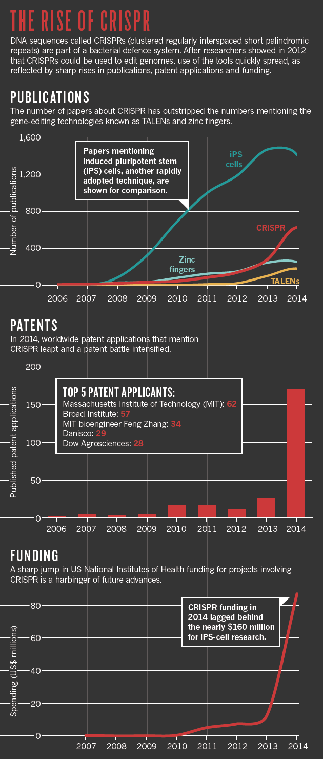
Publications: Scopus; Patents: The Lens; Funding: NIH RePORTER.

Researchers have traditionally relied heavily on model organisms such as mice and fruit flies, partly because they were the only species that came with a good tool kit for genetic manipulation. Now CRISPR is making it possible to edit genes in many more organisms. In April, for example, researchers at the Whitehead Institute for Biomedical Research in Cambridge, Massachusetts, reported using CRISPR to study Candida albicans, a fungus that is particularly deadly in people with weakened immune systems, but had been difficult to genetically manipulate in the lab3. Jennifer Doudna, a CRISPR pioneer at the University of California, Berkeley, is keeping a list of CRISPR-altered creatures. So far, she has three dozen entries, including disease-causing parasites called trypanosomes and yeasts used to make biofuels.

Yet the rapid progress has its drawbacks. “People just don’t have the time to characterize some of the very basic parameters of the system,” says Bo Huang, a biophysicist at the University of California, San Francisco. “There is a mentality that as long as it works, we don’t have to understand how or why it works.” That means that researchers occasionally run up against glitches. Huang and his lab struggled for two months to adapt CRISPR for use in imaging studies. He suspects that the delay would have been shorter had more been known about how to optimize the design of guide RNAs, a basic but important nuance.

By and large, researchers see these gaps as a minor price to pay for a powerful technique. But Doudna has begun to have more serious concerns about safety. Her worries began at a meeting in 2014, when she saw a postdoc present work in which a virus was engineered to carry the CRISPR components into mice. The mice breathed in the virus, allowing the CRISPR system to engineer mutations and create a model for human lung cancer4. Doudna got a chill; a minor mistake in the design of the guide RNA could result in a CRISPR that worked in human lungs as well. “It seemed incredibly scary that you might have students who were working with such a thing,” she says. “It’s important for people to appreciate what this technology can do.”

Andrea Ventura, a cancer researcher at Memorial Sloan Kettering Cancer Center in New York and a lead author of the work, says that his lab carefully considered the safety implications: the guide sequences were designed to target genome regions that were unique to mice, and the virus was disabled such that it could not replicate. He agrees that it is important to anticipate even remote risks. “The guides are not designed to cut the human genome, but you never know,” he says. “It’s not very likely, but it still needs to be considered.”

Editing out disease

Last year, bioengineer Daniel Anderson of the Massachusetts Institute of Technology in Cambridge and his colleagues used CRISPR in mice to correct a mutation associated with a human metabolic disease called tyrosinaemia5. It was the first use of CRISPR to fix a disease-causing mutation in an adult animal — and an important step towards using the technology for gene therapy in humans (see ‘A brief history of CRISPR’).

The idea that CRISPR could accelerate the gene-therapy field is a major source of excitement in scientific and biotechnology circles. But as well as highlighting the potential, Anderson’s study showed how far there is to go. To deliver the Cas9 enzyme and its guide RNA into the target organ, the liver, the team had to pump large volumes of liquid into blood vessels — something that is not generally considered feasible in people. And the experiments corrected the disease-causing mutation in just 0.4% of the cells, which is not enough to have an impact on many diseases.

Over the past two years, a handful of companies have sprung up to develop CRISPR-based gene therapy, and Anderson and others say that the first clinical trials of such a treatment could happen in the next one or two years. Those first trials will probably be scenarios in which the CRISPR components can be injected directly into tissues, such as those in the eye, or in which cells can be removed from the body, engineered in the lab and then put back. For example, blood-forming stem cells might be corrected to treat conditions such as sickle-cell disease or β-thalassaemia. It will be a bigger challenge to deliver the enzyme and guide RNA into many other tissues, but researchers hope that the technique could one day be used to tackle a wider range of genetic diseases.

Yet many scientists caution that there is much to do before CRISPR can be deployed safely and efficiently. Scientists need to increase the efficiency of editing, but at the same time make sure that they do not introduce changes elsewhere in the genome that have consequences for health. “These enzymes will cut in places other than the places you have designed them to cut, and that has lots of implications,” says Haber. “If you’re going to replace somebody’s sickle-cell gene in a stem cell, you’re going to be asked, ‘Well, what other damage might you have done at other sites in the genome?’”

Keith Joung, who studies gene editing at Massachusetts General Hospital in Boston, has been developing methods to hunt down Cas9’s off-target cuts. He says that the frequency of such cuts varies widely from cell to cell and from one sequence to another: his lab and others have seen off-target sites with mutation frequencies ranging from 0.1% to more than 60%. Even low-frequency events could potentially be dangerous if they accelerate a cell’s growth and lead to cancer, he says.

With so many unanswered questions, it is important to keep expectations of CRISPR under control, says Katrine Bosley, chief executive of Editas, a company in Cambridge, Massachusetts, that is pursuing CRISPR-mediated gene therapy. Bosley is a veteran of commercializing new technologies, and says that usually the hard part is convincing others that an approach will work. “With CRISPR it’s almost the opposite,” she says. “There’s so much excitement and support, but we have to be realistic about what it takes to get there.”

CRISPR on the farm

While Anderson and others are aiming to modify DNA in human cells, others are targeting crops and livestock. Before the arrival of gene-editing techniques, this was generally done by inserting a gene into the genome at random positions, along with sequences from bacteria, viruses or other species that drive expression of the gene. But the process is inefficient, and it has always been fodder for critics who dislike the mixing of DNA from different species or worry that the insertion could interrupt other genes. What is more, getting genetically modified crops approved for use is so complex and expensive that most of those that have been modified are large commodity crops such as maize (corn) and soya beans.

Illustration by Sébastien Thibault

With CRISPR, the situation could change: the ease and low cost may make genome editing a viable option for smaller, speciality crops, as well as animals. In the past few years, researchers have used the method to engineer petite pigs and to make disease-resistant wheat and rice. They have also made progress towards engineering dehorned cattle, disease-resistant goats and vitamin-enriched sweet oranges. Doudna anticipates that her list of CRISPR-modified organisms will grow. “There’s an interesting opportunity to consider doing experiments or engineering pathways in plants that are not as important commercially but are very interesting from a research perspective — or for home vegetable gardens,” she says.

CRISPR’s ability to precisely edit existing DNA sequences makes for more-accurate modifications, but it also makes it more difficult for regulators and farmers to identify a modified organism once it has been released. “With gene editing, there’s no longer the ability to really track engineered products,” says Jennifer Kuzma, who studies science policy at North Carolina State University in Raleigh. “It will be hard to detect whether something has been mutated conventionally or genetically engineered.”

That rings alarm bells for opponents of genetically modified crops, and it poses difficult questions for countries trying to work out how to regulate gene-edited plants and animals. In the United States, the Food and Drug Administration has yet to approve any genetically modified animal for human consumption, and it has not yet announced how it will handle gene-edited animals.

Under existing rules, not all crops made by genome editing would require regulation by the US Department of Agriculture (see Nature 500, 389390; 2013). But in May, the agriculture department began to seek input on how it can improve regulation of genetically modified crops — a move that many have taken as a sign that the agency is re-evaluating its rules in light of technologies such as CRISPR. “The window has been cracked,” says Kuzma. “What goes through the window remains to be seen. But the fact that it’s even been cracked is pretty exciting.”

Engineered ecosystems

Beyond the farm, researchers are considering how CRISPR could or should be deployed on organisms in the wild. Much of the attention has focused on a method called gene drive, which can quickly sweep an edited gene through a population. The work is at an early stage, but such a technique could be used to wipe out disease-carrying mosquitoes or ticks, eliminate invasive plants or eradicate herbicide resistance in pigweed, which plagues some US farmers.

Usually, a genetic change in one organism takes a long time to spread through a population. That is because a mutation carried on one of a pair of chromosomes is inherited by only half the offspring. But a gene drive allows a mutation made by CRISPR on one chromosome to copy itself to its partner in every generation, so that nearly all offspring will inherit the change. This means that it will speed through a population exponentially faster than normal (see ‘Gene drive’) — a mutation engineered into a mosquito could spread through a large population within a season. If that mutation reduced the number of offspring a mosquito produced, then the population could be wiped out, along with any malaria parasites it is carrying.
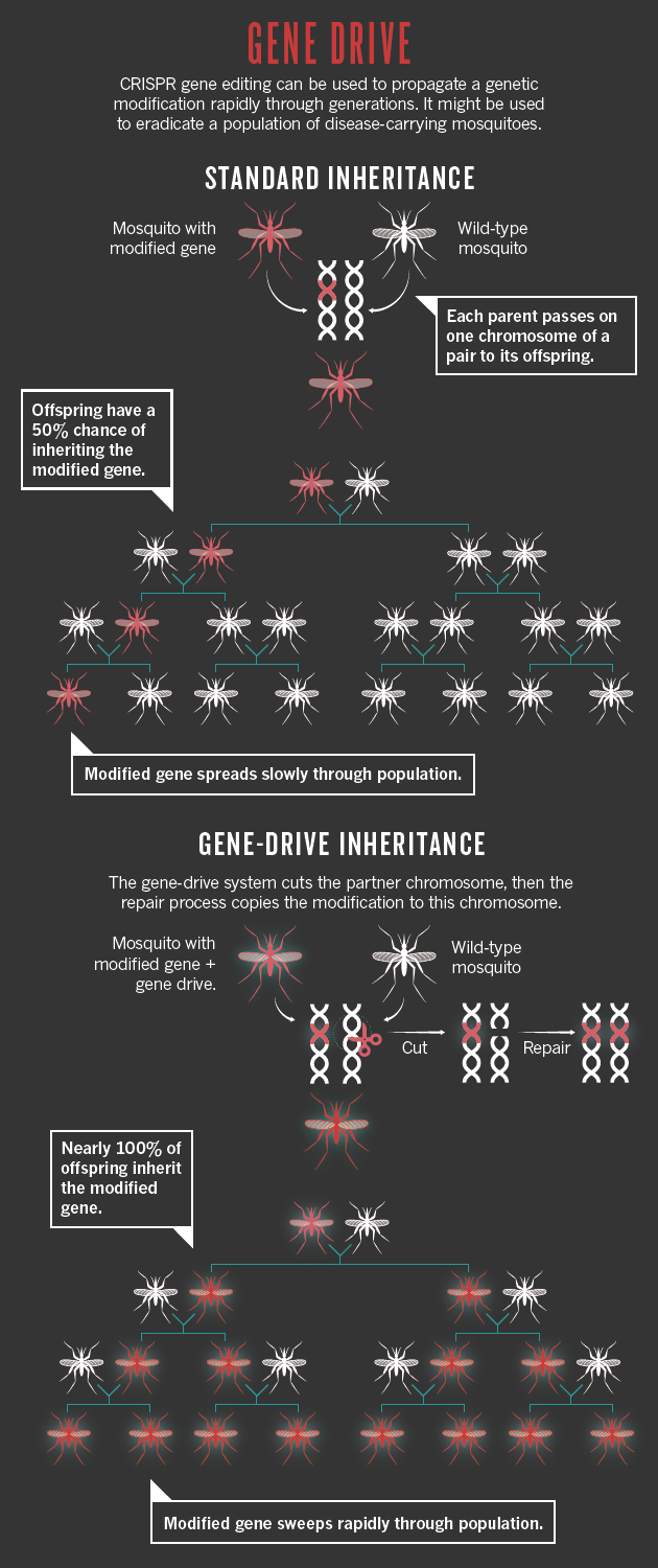
Publications: Scopus; Patents: The Lens; Funding: NIH RePORTER.

But many researchers are deeply worried that altering an entire population, or eliminating it altogether, could have drastic and unknown consequences for an ecosystem: it might mean that other pests emerge, for example, or it could affect predators higher up the food chain. And researchers are also mindful that a guide RNA could mutate over time such that it targets a different part of the genome. This mutation could then race through the population, with unpredictable effects.

“It has to have a fairly high pay-off, because it has a risk of irreversibility — and unintended or hard-to-calculate consequences for other species,” says George Church, a bioengineer at Harvard Medical School in Boston. In April 2014, Church and a team of scientists and policy experts wrote a commentary in Science6 warning researchers about the risks and proposing ways to guard against accidental release of experimental gene drives.

At the time, gene drives seemed a distant prospect. But less than a year later, developmental biologist Ethan Bier of the University of California, San Diego, and his student Valentino Gantz reported that they had designed just such a system in fruit flies7. Bier and Gantz had used three layers of boxes to contain their flies and adopted lab safety measures usually used for malaria-carrying mosquitoes. But they did not follow all the guidelines urged by the authors of the commentary, such as devising a method to reverse the engineered change. Bier says that they were conducting their first proof-of-principle experiments, and wanted to know whether the system worked at all before they made it more complex.

For Church and others, this was a clear warning that the democratization of genome editing through CRISPR could have unexpected and undesirable outcomes. “It is essential that national regulatory authorities and international organizations get on top of this — really get on top of it,” says Kenneth Oye, a political scientist at the Massachusetts Institute of Technology and lead author of the Science commentary. “We need more action.” The US National Research Council has formed a panel to discuss gene drives, and other high-level discussions are starting to take place. But Oye is concerned that the science is moving at lightning speed, and that regulatory changes may happen only after a high-profile gene-drive release.

The issue is not black and white. Micky Eubanks, an insect ecologist at Texas A&M University in College Station, says that the idea of gene drives shocked him at first. “My initial gut reaction was ‘Oh my god, this is terrible. It’s so scary’,” he says. “But when you give it more thought and weigh it against the environmental changes that we have already made and continue to make, it would be a drop in the ocean.”

Some researchers see lessons for CRISPR in the arc of other new technologies that prompted great excitement, concern and then disappointment when teething troubles hit. Medical geneticist James Wilson of the University of Pennsylvania in Philadelphia was at the centre of booming enthusiasm over gene therapy in the 1990s — only to witness its downfall when a clinical trial went wrong and killed a young man. The field went into a tailspin and has only recently begun to recover. The CRISPR field is still young, Wilson says, and it could be years before its potential is realized. “It’s in the exploration stage. These ideas need to ferment.”

Then again, Wilson has been bitten by the CRISPR bug. He says that he was sceptical of all the promises being made about it until his own lab began to play with the technique. “It’s ultimately going to have a role in human therapeutics,” he says. “It’s just really spectacular.”
FunCti.Oning · MEchA.NIC · ((GeArS) in · a · .Bio.Logi:CaL, StruC.Ture in · Natu:Re Cre;AtiNg,, Abso:Lute ,. Syn:CHroni:CiTY

functioningmbaktera

Sou:Rce : http://phys.org/news/2013-09-functioning-mechanical-gears-nature.html

Functioning ‘mechanical gears’ seen in nature for the first time

September 12, 2013
This image shows cog wheels connecting the hind legs of the plant hopper, Issus. Credit: Burrows/Sutton

a plant-hopping insect found in gardens across Europe – has hind-leg joints with curved cog-like strips of opposing ‘teeth’ that intermesh, rotating like mechanical gears to synchronise the animal’s legs when it launches into a jump.

The finding demonstrates that gear mechanisms previously thought to be solely man-made have an evolutionary precedent. Scientists say this is the “first observation of mechanical gearing in a “.

Through a combination of anatomical analysis and high-speed video capture of normal Issus movements, scientists from the University of Cambridge have been able to reveal these functioning natural gears for the first time. The findings are reported in the latest issue of the journal Science.

The gears in the Issus hind-leg bear remarkable engineering resemblance to those found on every bicycle and inside every car gear-box.

Each gear tooth has a rounded corner at the point it connects to the gear strip; a feature identical to man-made gears such as bike gears – essentially a shock-absorbing mechanism to stop teeth from shearing off.

The gear teeth on the opposing hind-legs lock together like those in a car gear-box, ensuring almost complete synchronicity in leg movement – the legs always move within 30 ‘‘ of each other, with one microsecond equal to a millionth of a second.

This is critical for the powerful jumps that are this insect’s primary mode of transport, as even miniscule discrepancies in synchronisation between the velocities of its legs at the point of propulsion would result in “yaw rotation” – causing the Issus to spin hopelessly out of control.

Functioning 'mechanical gears' seen in nature for the first time
Photograph of an Issus nymph. Credit: Malcolm Burrows

“This precise synchronisation would be impossible to achieve through a nervous system, as would take far too long for the extraordinarily tight coordination required,” said lead author Professor Malcolm Burrows, from Cambridge’s Department of Zoology.

“By developing mechanical gears, the Issus can just send nerve signals to its muscles to produce roughly the same amount of force – then if one leg starts to propel the jump the gears will interlock, creating absolute.

n Issus, the skeleton is used to solve a complex problem that the brain and nervous system can’t,” said Burrows. “This emphasises the importance of considering the properties of the skeleton in how movement is produced.”

“We usually think of gears as something that we see in human designed machinery, but we’ve found that that is only because we didn’t look hard enough,” added co-author Gregory Sutton, now at the University of Bristol.

“These gears are not designed; they are evolved – representing high speed and precision machinery evolved for synchronisation in the animal world.”

Interestingly, the mechanistic gears are only found in the insect’s juvenile – or ‘nymph’ – stages, and are lost in the final transition to adulthood. These transitions, called ‘molts’, are when animals cast off rigid skin at key points in their development in order to grow.

It’s not yet known why the Issus loses its hind-leg gears on reaching adulthood. The scientists point out that a problem with any gear system is that if one tooth on the gear breaks, the effectiveness of the whole mechanism is damaged. While gear-teeth breakage in nymphs could be repaired in the next molt, any damage in adulthood remains permanent.

Spontaneous jump of a nymph viewed from the side. The images were captured at a rate of 5,000 images s-1 and with an exposure time of 0.03 ms and are replayed at 30 frames s-1 . Credit: Malcolm Burrows

It may also be down to the larger size of adults and consequently their ‘trochantera’ – the insect equivalent of the femur or thigh bones. The bigger adult trochantera might allow them to can create enough friction to power the enormous leaps from leaf to leaf without the need for intermeshing gear teeth to drive it, say the scientists.

Each gear strip in the juvenile Issus was around 400 micrometres long and had between 10 to 12 teeth, with both sides of the gear in each leg containing the same number – giving a gearing ratio of 1:1.

Unlike man-made gears, each gear tooth is asymmetrical and curved towards the point where the cogs interlock – as man-made gears need a symmetric shape to work in both rotational directions, whereas the Issus gears are only powering one way to launch the animal forward.

While there are examples of apparently ornamental cogs in the animal kingdom – such as on the shell of the cog wheel turtle or the back of the wheel bug – gears with a functional role either remain elusive or have been rendered defunct by evolution.

The Issus is the first example of a natural cog mechanism with an observable function, say the scientists.